 |
| |
 |
 |
|
Lumps or masses in the breast are not unusual, and most of them are not cancerous.
In fact, 8 out of 10 lumps are not cancerous. In order to avoid unnecessary
surgery and anesthesia, these lumps or masses can be sampled by using a needle
and image guidance – either by stereotactic or ultrasound guidance.
 During a stereotactic biopsy procedure, the patient lies on her abdomen on
a specially designed exam table. An opening in the table allows access to the
breast. The table is raised and the biopsy is done from below the table. Using
a local anesthetic, the radiologist or surgeon makes a small opening in the
skin. A sterile biopsy needle is placed into the breast tissue area to be biopsied.
Computerized images help confirm the needle placement using digital imaging.
Tissue samples are taken through the needle using a special vacuum-assisted
device called a mammotome. It is common to take multiple tissue samples in
one very small location. This part of the biopsy takes approximately 15
minutes. After the biopsy samples are taken, a small titanium clip is
usually inserted into the area to mark the biopsy site. A
mammogram
is then taken of the effected breast to show placement of
the clip. Upon completion, sterile strips and a small dressing are applied
to the skin, as well as a small cold pack. A mammogram is usually taken after
the procedure Results are available in 24 to 48 hours.
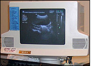
During this procedure, the patient lies on her back, on the table. Using a
local anesthetic, the radiologist makes a small opening in the skin. A sterile
biopsy needle is placed into the breast tissue area to be biopsied. The radiologist
uses ultrasound to find the area
in question and guide the needle to that spot. Tissue samples are taken utilizing
a special needle or a vacuum-assisted device called a mammotome system. It
is common to take multiple tissue samples in one very small location. This
part of the biopsy takes approximately 15 minutes. Sometimes it is necessary
to insert a special clip into the biopsied area to mark the site that samples
were taken. If a clip was inserted, a
mammogram is
usually performed to show the placement of the clip. Upon completion, sterile
strips and a small dressing are applied to the skin, as well as a small cold
pack. Results are available in 24 to 48 hours.
|
 |

For
More Information
(419) 226-4500 |
| Fact:
|
There are over two million breast cancer survivors in the United States today.
-Source: National Association for Breast Cancer
|
|

